May 2023
Myelolipoma
Reviewer(s): Dharam Ramnani, MD | Last Update: 5/24/2023
Myelolipoma is a benign tumor-like lesion with mature fat and normal bone marrow elements, usually seen in the adrenals in individuals > 40 years. It has been associated with hormonally active adrenal tumors/lesions. Rare cases are in extraadrenal locations.
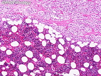
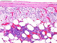
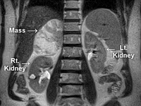
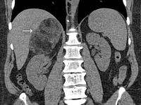
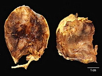
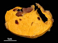
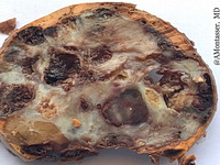
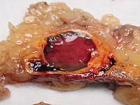
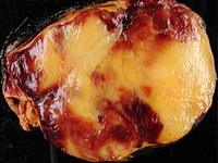
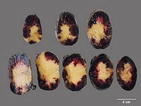
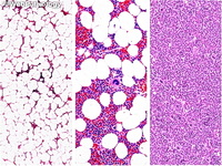
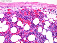
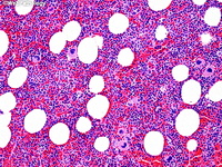
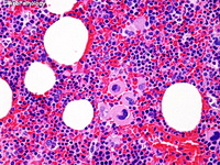
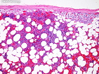
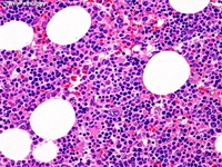
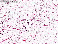
Most tumors are small, non-functioning, asymptomatic and often detected incidentally on imaging studies or surgery. Some reach large size and may spontaneously rupture producing massive retroperitoneal hemorrhage.Grossly, it is a soft, well-circumscribed mass ranging in size from 3 to 8 cm but may reach 30 cm or more. The cut surface shows an admixture of yellow adipose tissue and gelatinous, reddish-brown areas (marrow elements). Microscopically, there are varying proportions of mature fat and normal trilineage hematopoietic elements with increased number of megakaryocytes.Several cases with t(3;21)(q25;p11) have been documented. Both hematopoietic and adipose tissue are derived from the same clone, suggesting that it is a true neoplasm. Pre-operative diagnosis can be made with imaging studies and FNA. Small asymptomatic lesions can be left alone. Large symptomatic masses are resected.
Most tumors are small, non-functioning, asymptomatic and often detected incidentally on imaging studies or surgery. Some reach large size and may spontaneously rupture producing massive retroperitoneal hemorrhage.Grossly, it is a soft, well-circumscribed mass ranging in size from 3 to 8 cm but may reach 30 cm or more. The cut surface shows an admixture of yellow adipose tissue and gelatinous, reddish-brown areas (marrow elements). Microscopically, there are varying proportions of mature fat and normal trilineage hematopoietic elements with increased number of megakaryocytes.Several cases with t(3;21)(q25;p11) have been documented. Both hematopoietic and adipose tissue are derived from the same clone, suggesting that it is a true neoplasm. Pre-operative diagnosis can be made with imaging studies and FNA. Small asymptomatic lesions can be left alone. Large symptomatic masses are resected.


