
Myelolipoma : Imaging

slide 4 of 17


Comments:
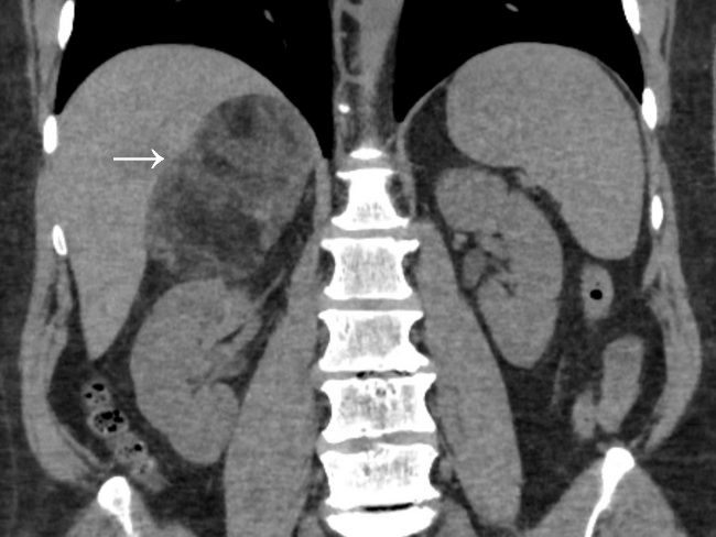
Imaging: This coronal non-contrast CT image is from the same case as the previous MRI. Sixty year-old male with right flank pain was found to have a right retroperitoneal mass on ultrasonography. The image shows an oval, well-circumscribed right suprarenal mass with mixed densities (gross fat plus soft tissue densities). The mass has displaced the right kidney downwards. The imaging findings are classic for myelolipoma. Case courtesy of Mostafa El-Feky, Radiopaedia.org. From the case rID: 65240
slide 4 of 17