
Myeloma : M Component


Comments:
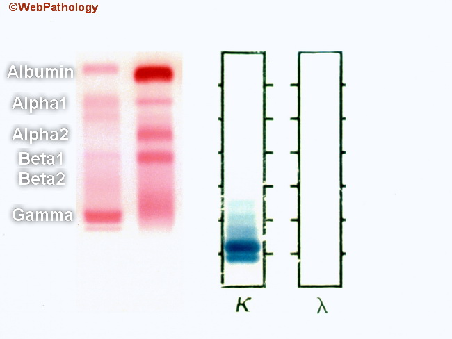
This image shows serum protein electrophoresis (left half; red bands) and urine protein electrophoresis (right half; blue bands) from a patient with plasma cell myeloma. The right lane in serum protein electrophoresis (red bands) is a normal control showing a thick albumin band at the top followed by alpha1, alpha2, beta1, beta2 (indistinct; merges with gamma), and gamma bands. The left lane shows serum from a patient with plasma cell myeloma. Note the sharply defined band in the gamma region at the bottom. Also note the reduced levels of albumin and other polyclonal immunoglobulins. With immunofixation technique, the M component was identified as IgG kappa. The right half of this image depicts urine protein electrophoresis from the same patient. It shows the presence of free kappa light chains (Bence-Jones proteins) in urine. The free light chains are reabsorbed by proximal renal tubules and they may be absent from urine if the renal function is preserved. Plasma cell myeloma patients usually show increased levels of serum immunoglobulins (>3 gm/dl) and/or Bence-Jones proteins in urine (> 6 mg/dl). The M component is usually IgG (about 50% of cases) followed by IgA (about 20% of cases) type. Myelomas producing M component of IgM, IgD, and IgE types are rare. About 20% of myeloma cases show only free light chains, usually kappa subtype (Bence-Jones proteins) in urine and no M component in the serum. About 3% of myelomas are non-secretory; therefore, the absence of M component in the serum or urine does not rule out myeloma. Excessive levels of immunoglobulins produce hyperviscosity syndrome in about 7% of patients.