
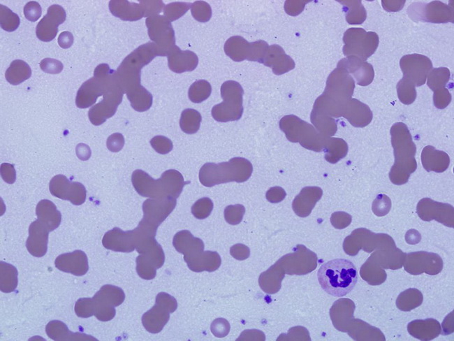
Myeloma : Rouleaux formation


Comments:
High levels of monoclonal immunoglobulins in patients with myeloma cause the red cells in peripheral blood smears to stick to one another in a linear fashion - a phenomenon labeled rouleaux formation. The high levels of M component also cause bluish staining of the background on Wright-Giemsa stain as seen in this image. Rouleaux formation is not specific for plasma cell disorders. Any condition that raises Ig levels, such as lupus, chronic infections, and HIV can cause rouleaux formation. A few Plasma cells may be seen in peripheral blood in about 15% of myeloma cases. Marked plasmacytosis is seen in more advanced stages of the disease termed plasma cell leukemia. Image courtesy of: Jian-Hua Qiao, MD, FCAP, Los Angeles, CA, USA.