
Aneurysmal Bone Cyst : Case 3

slide 11 of 40


Comments:
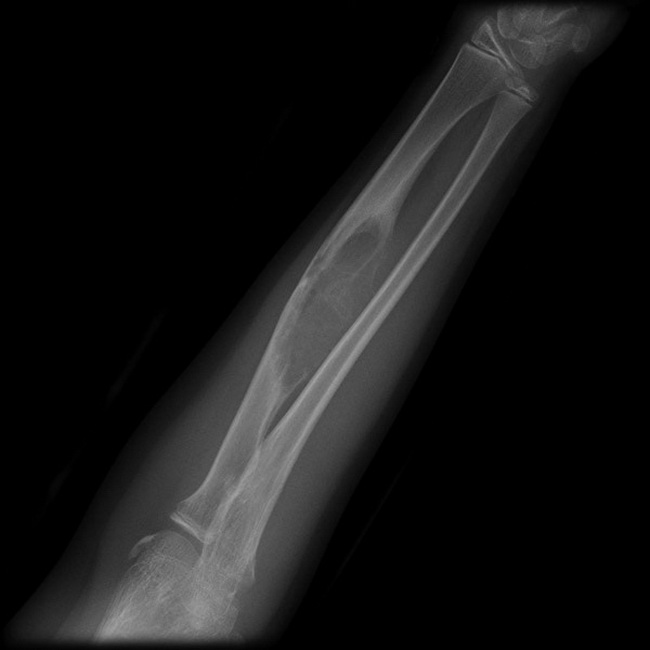
Case History: Child with a painful left forearm. Radiograph shows an expansile bubbly lesion located eccentrically in mid-radial diaphysis. The lesion has an aggressive appearance with irregular margins, loss of cortex, no convincing matrix mineralization and no soft tissue extension. Bone scans demonstrated increased uptake. MRI showed multiple septa and fluid-fluid levels (see next image), which although non-specific suggested the diagnosis of an aneurysmal bone cyst. This was confirmed on histology following resection and grafting. Case courtesy of Assoc Prof Frank Gaillard, Radiopaedia.org. From the case rID: 5866
slide 11 of 40