
Aneurysmal Bone Cyst : Case 3

slide 12 of 40


Comments:
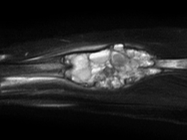
Case History: Child with a painful left forearm (same case as the previous image). MRI (sagittal T2 image) demonstrates an expansile mid-radial mass containing multiple septa and fluid-fluid levels, which although non specific suggest the diagnosis of an aneurysmal bone cyst (ABC). This was confirmed on histology following resection and grafting. Fluid-fluid levels are seen in ABC and telangiectatic osteosarcoma. Case courtesy of Assoc Prof Frank Gaillard, Radiopaedia.org. From the case rID: 5866
slide 12 of 40