
Aneurysmal Bone Cyst : Case 2

slide 10 of 40


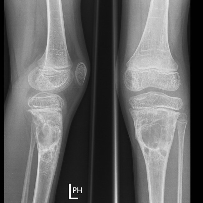
Comments:
Case History: Male child who presented with pain around left knee. X-rays demonstrate an expansile, multiloculated lucency in the metaphysis of upper tibia. There is no fracture although the posterior cortex appears deficient. The lesion does not transgress the growth plate. This patient went on to have further imaging and eventually had the lesion operated upon, and confirmed to be an aneurysmal bone cyst. Region around the knee (distal femur and proximal tibia) is the most common site for aneurysmal bone cyst; however, any portion of the skeleton may be involved. Radiologic features of ABCs are discussed in images 3 and 4. Case courtesy of Assoc Prof Frank Gaillard, Radiopaedia.org. From the case rID: 19346
slide 10 of 40