Testicular Adnexa : Benign Tumors
Reviewer(s): Dharam Ramnani, MD
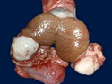
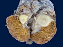
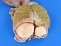
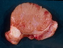
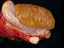
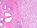
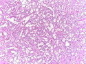
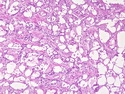
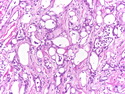
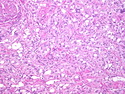
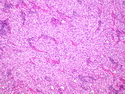
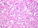
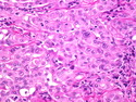
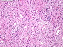
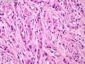
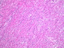
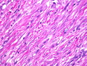
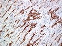
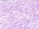
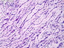
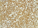
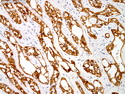
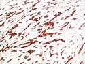
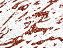
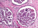
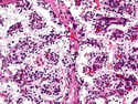
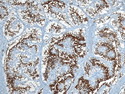
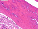
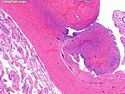
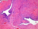
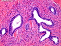
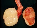
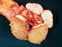
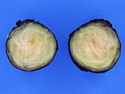
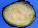
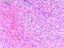
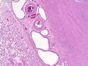
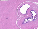
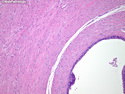
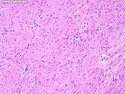
Benign tumors of testicular adnexa include adenomatoid tumor (most common tumor of the epididymis), lipoma (most common tumor of the spermatic cord), papillary cystadenoma of the epididymis, leiomyoma, rhabdomyoma, hemangioma, lymphangioma, aggressive angiomyxoma and paraganglioma. Fibrous pseudotumor and smooth muscle hyperplasia of the testicular adnexa are entities reported in this region that mimic neoplasia clinically. Ovarian-type epithelial tumors (serous, mucinous, Brenner, endometrioid, and clear cell types) have also been described in paratesticular tissues.