
Adenoid Cystic CA : Microscopic


Comments:
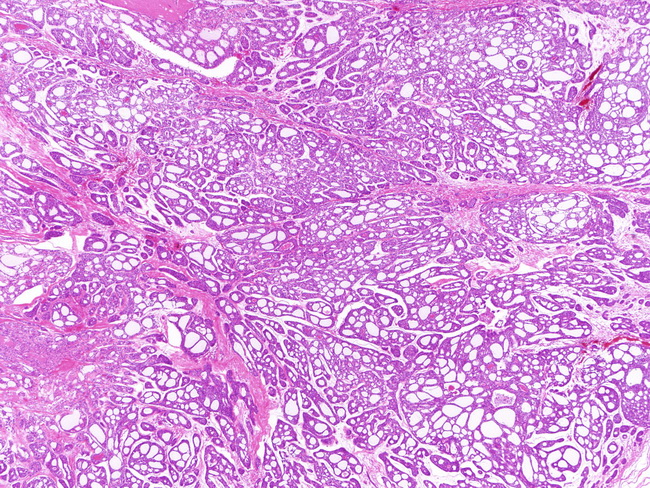
GROSS: It is solid, yellow-tan with ill-defined margins due to infiltrative growth pattern. Rare cases are well-circumscribed. MICROSCOPIC: Adenoid cystic carcinoma shows an admixture of many architectural patterns, including cribriform, tubular, and solid growth with a myxohyaline stroma. There are two cell types � abundant basaloid cells with myoepithelial differentiation and eosinophilic epithelial cells with ductal differentiation. The cribriform areas (as seen in this low magnification view) are almost always present, even if focally. They consist of variable-sized, smooth contoured lobules or islands of uniform basaloid cells punctuated by round rigid gland-like spaces. These spaces are not true glandular lumens but represent stromal invaginations (pseudocysts). They are filled with eosinophilic material (PAS-positive, diastase-resistant; reduplicated basal lamina) or basophilic myxoid mucinous material (Alcian blue positive). True glandular structures lined by low cuboidal cells with eosinophilic cytoplasm are also present. Image courtesy of: @Patholwalker