
Adenoid Cystic CA : Intro & Clinical


Comments:
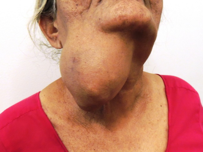
INTRODUCTION: Adenoid cystic carcinoma is a slow-growing but highly aggressive tumor with a relentless course and poor long-term prognosis. It is one of the common tumors of salivary glands, occurring more frequently in minor salivary glands. In the parotid gland, it is seen less commonly than mucoepidermoid and acinic cell carcinomas. Besides salivary glands, it also occurs in other exocrine glands in sites such as breast, lung, trachea, paranasal sinuses, eye (lacrimal glands), cervix and vulva (Bartholin glands), prostate, gastrointestinal tract, and skin. Most cases are diagnosed between 4th and 6th decades of life. Pediatric cases have been recorded. There is slight female preponderance (F:M = 3:2). The tumor originates from one of the intercalated duct cells that is capable of differentiating along both epithelial and myoepithelial lines. CLINICAL PRESENTATION: Early lesions of salivary glands present as slow-growing painless mass of oral cavity or face. More advances cases can present with pain, tenderness and facial nerve palsy due to its propensity for perineural invasion. Larger tumors are often fixed to the overlying skin or deeper tissues. Palatal tumors can ulcerate. The tumor can also infiltrate into adjacent bony structures. CASE HISTORY: The patient was a 68 y/o female who presented with a 1-yr history of a slowly growing mass on the right side of her neck as well as change in her voice. The tumor had shown rapid growth in the last 2 months before presentation. It was hard in consistency, non-tender and fixed to the overlying skin. MRI scan showed a multilobulated mass measuring 11.6 x 6.5 x 6 cm involving right oropharyngeal and parapharyngeal space. The mass appeared to be starting from oral cavity and extending caudally up to C5 level . The carotid and jugular vessels were displaced by the mass. Microscoic examination confirmed lesion as adenoid cystic carcinoma. Onset of rapid growth in a previously slow-growing tumor is often a sign of de-differentiation. Case courtesy of: Dr. Sanjay D. Deshmukh (Professor of Pathology) & Dr. J. M. Gadekar (Prof.and Head, Surgery Dept.), Dr. Vithalrao Vikhe Patil Medical Foundation�s College & Hospitals, Ahmednagar, India.