
SMZL : Morphology
Home
 Hematopathology
Mature B-cell Neoplasms - Part I
Splenic Marginal Zone Lymphoma
SMZL : Morphology
Hematopathology
Mature B-cell Neoplasms - Part I
Splenic Marginal Zone Lymphoma
SMZL : Morphology

slide 4 of 24


Comments:
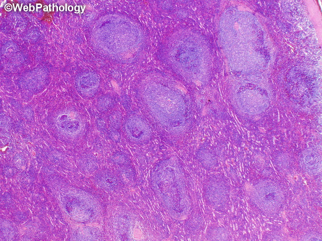
Splenic Marginal Zone Lymphoma (SMZL) - Morphology: The tumor is centered around the white pulp which shows replacement of preexisting lymphoid follicles by micronodular tumoral infiltrates. At low magnification, one sees marginal zone differentiation with a biphasic appearance. The interior of the follicles show darker small lymphocytes with scant cytoplasm and round or oval nuclei. They merge with larger pale-staining cells in the marginal zone that have more abundant cytoplasm and irregular nuclei. A few regressed or reactive germinal centers may be seen within some of the follicles.
slide 4 of 24