
Calcifying Fibrous Tumor : Gross & Microscopic


Comments:
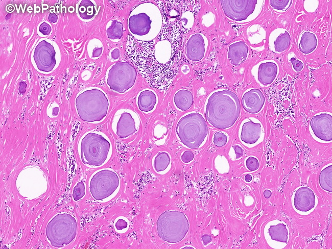
Gross Pathology: Calcifying fibrous tumor (CFT) is a solid, well-circumscribed, lobulated mass with firm consistency. It has a wide size range from 3 cm to > 15 cm; most measure 3 to 5 cm at the time of excision. It has a homogenous grayish-white appearance and cuts with a gritty sensation due to calcification. Microscopic Features: CFT is circumscribed, unencapsulated mass located in the subcutaneous or deep soft tissues. It is hypocellular and consists mainly of dense, hyalinized eosinophilic collagenous matrix containing psammomatous calcifications. In some cases, calcification may be extensive and make up bulk of the tumor. Bland fibroblastic and myofibroblastic spindle cells are present in the stroma. Scattered lymphoplasmacytic cells and rare germinal centers may be seen. Immunohistochemistry: The spindle cells are CD34-positive and there is variable expression of muscle markers (SMA, MSA, & desmin). These markers are never diffusely or strongly positive. ALK is consistently negative.