
Calcifying Fibrous Tumor : Differential Diagnosis


Comments:
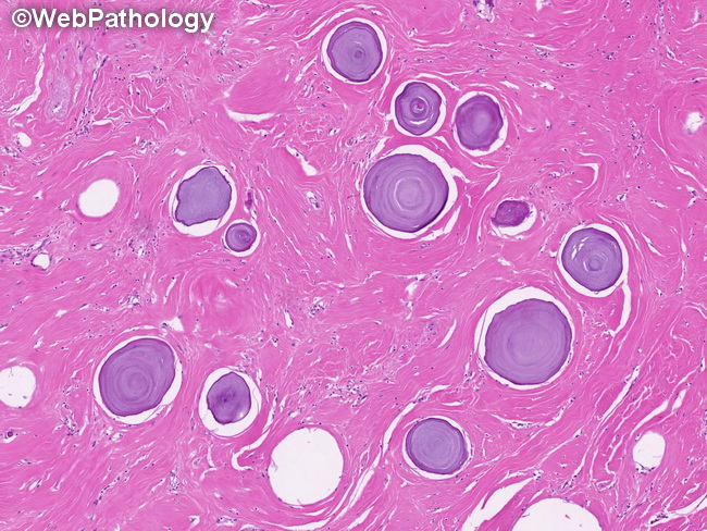
Differential Diagnosis: The differential diagnosis of calcifying fibrous tumor (CFT) includes several benign, reactive, and neoplastic fibroblastic proliferations. These entities and their distinguishing features are:Inflammatory Myofibroblastic Tumor: greater cellularity, less hyalinization, no calcification, ALK positive (including ALK gene rearrangements).Reactive Nodular Fibrous Pseudotumor: less cellular than CFT, no inflammatory infiltrate, no calcifications, positive for desmin, actin, & CD117.Desmoid-type Fibromatosis: poorly circumscribed with infiltrative growth, greater cellularity, fascicular growth, rare calcifications.Desmoplastic Fibroblastoma: more common in adults with a male predilection; fibrous mass in the subcutaneous tissues or skeletal muscles; consists of dense collagenous stroma but lacks calcifications of CFT.Nodular Fasciitis: tissue culture-like spindle cells, myxoid matrix, rare/no calcifications. Fibroma of Tendon Sheath: Distal extremity location, dense eosinophilic collagen but with foci of increased cellularity, elongated slit-like spaces, no calcifications.Calcifying Aponeurotic Fibroma: Location in hands and feet, poorly-circumscribed, calcifications and chondroid metaplasia, multinucleated giant cells.Amyoidoma: Dense eosinophilic deposits that are Congo-Red positive.