
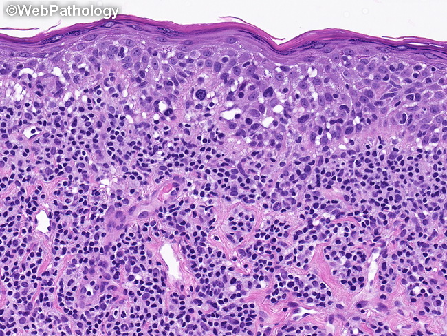
Mycosis Fungoides : Plaque Stage


Comments:
Mycosis fungoides (MF) - Plaque Stage: Large atypical lymphocytes with irregular nuclei are more numerous than in the late patch stage and constitute a significant proportion of the infiltrate. They are present singly or in clusters within the epidermis, sometimes obscuring the dermal-epidermal junction. Intraepidermal vesicles with atypical lymphocytes (Pautrier microabscess) more frequent than in the patch stage. Several Pautrier microabscesses are seen in this image. Epidermotropism may be inconspicuous if the biopsy is taken after topical treatment. In the dermis, the lymphocytes are present in a perivascular distribution as well as in between reticular dermal collagen bundles. In addition to the atypical lymphoid cells, the dermal infiltrate consists of eosinophils, plasma cells, and histiocytes. Papillary dermis shows increased fibrosis.