
Mesenchymal Chondrosarcoma : Differential


Comments:
Differential Diagnosis (continued from previous image): Mesenchymal Chondrosarcoma (MC) vs Ewing Sarcoma (ES): Small undifferentiated cells of MC have more variability in nuclear size/shape than those in ES. CD99 and NKX2-2 (a nuclear marker expressed by 100% of ES) are also seen in MC. Features supporting ES include: absence of neoplastic cartilage; absence of spindle cells; presence of FLI1 expression (seen in 70% of ES; has 90% specificity); molecular evidence of EWSR1; absence of HEY1-NCOA2 fusion; absence of SOX9 expression
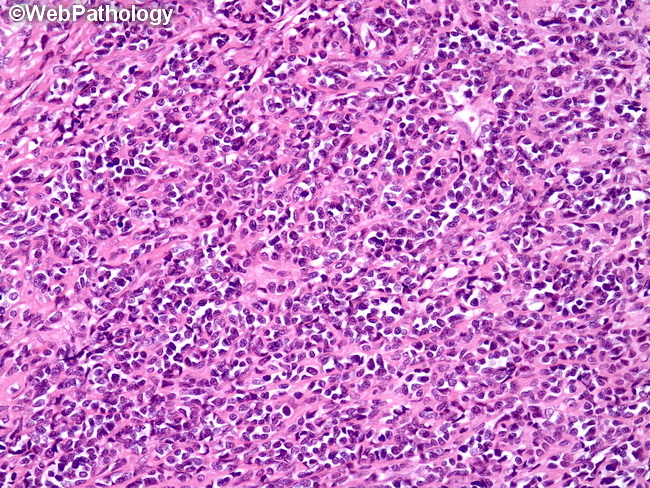
Mesenchymal Chondrosarcoma vs Synovial Sarcoma (SS): SS may show hemangiopericytoma-like vasculature and small foci of metaplastic cartilage; however, calcification or ossification is rare. Cytokeratin or EMA may reveal foci of epithelial differentiation in SS. The definitive answer will come from molecular studies (FISH test using SSX1 probe). This image shows sheets of undifferentiated small blue cells from a case of MC.