
Chordoid Meningioma

slide 48 of 60


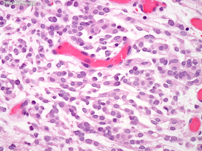
Comments:
Meningothelial cells arranged in chords displaying fused cytoplasmic membranes and indistinguishable cellular borders. The nuclei look bland; however this is by definition a Grade II meningioma. Despite the benign-looking features, chordoid meningiomas tend to recur, especially after subtotal resection.
slide 48 of 60