
Angiomatous Meningioma

slide 39 of 60


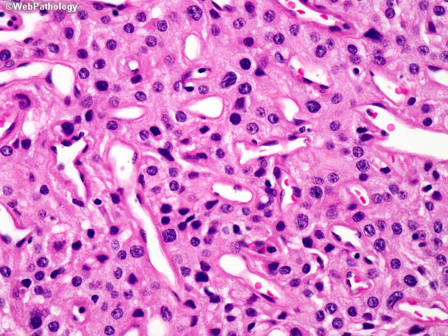
Comments:
Higher magnification of the previous picture illustrating the uniform evenly spaced meningothelial cells with indistinct borders, bland nuclear cytologic features, fine open chromatin, and inconspicuous nucleoli. Occasional hyperchromatic nuclei are present. Multiple ovoid to round shaped vascular spaces lined by a monolayer of endothelial cells can be noted.
slide 39 of 60