
Meningothelial Meningioma

slide 15 of 60


Comments:
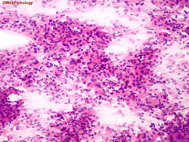
Smears often help in making the diagnosis of meningioma during intraoperative consultations. This photomicrograph shows sheets of monomorphic meningothelial cells suggesting syncytia formation. The cytoplasm is pink and ample; the nuclei are round to ovoid with fine grey chromatin.
slide 15 of 60