
Lipofibromatosis : Clinical Features


Comments:
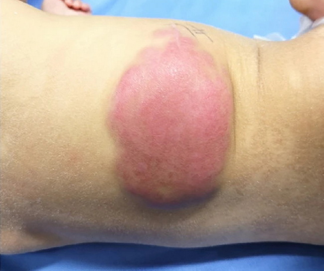
Clinical Features: Lipofibromatosis (LF) usually presents within 2 yrs. of age (range - birth to about 12 years) with a male predilection. About 20% of cases are congenital. The most common location is subcutis or skeletal muscles of the extremities (80% of cases involve hands, arm, leg, feet). Other sites include trunk, and head and neck region (tongue, mandible, maxilla, mastoid process, orbit). LF presents as a slow growing, solitary, ill-defined firm mass that may produce enlargement of the affected limb segment. It is usually asymptomatic but may cause mild pain, tenderness or functional disturbances as it infiltrates around blood vessels and nerves in later stages. Involvement of nearby joint capsule can limit movement. Lesions near mandible, maxilla, or mastoid frequently extend into the bone and mimic desmoplastic fibroma of bone. Case History: The image shows lipofibromatosis presenting as a 11 cm x 6 cm erythematous lesion on the back of a 6 month old female, mimicking infantile hemangioma. The lesion was first noted a few days after birth and slowly enlarged to its current size. The location is somewhat atypical; however, the diagnosis of lipofibromatosis was confirmed after resection. Image source: Li, Z., Zou, Y., Xu, G. et al. Giant dorsal lipofibromatosis in an infant: a case report. BMC Pediatr 22, 59 (2022). https://doi.org/10.1186/s12887-022-03130-7; cropped from original and used under Creative Commons - Attribution 4.0 International - CC BY 4.0