
Mesenchymal Chondrosarcoma : HEY1-NCOA2 Fusion


Comments:
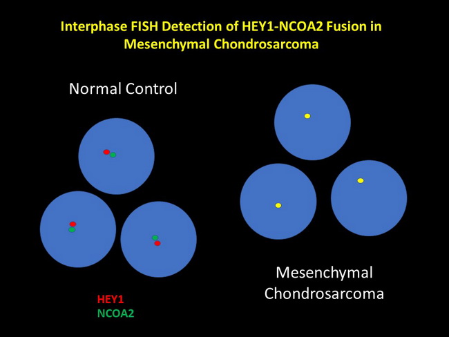
Molecular Pathogenesis: A recurrent t(8;8)(q21.1;q13.3) translocation resulting in HEY1-NCOA2 gene fusion is seen in 90% of mesenchymal chondrosarcomas. This can be detected by RT-PCR or FISH. Interpretation of FISH images can be challenging since both genes are located close to one another on chromosome 8. Rare cases show t(1;5) resulting in IRF2BP2-CDX1 fusion. IDH1 and IDH2 mutations are not found in mesenchymal chondrosarcomas - an important distinguishing feature from conventional and dedifferentiated chondrosarcomas. This schematic diagram illustrates detection of HEY1-NCOA2 fusion in mesenchymal chondrosarcoma by dual color FISH. Normal signal pattern (separate red and green dots placed close together) is seen on the left. Mesenchymal chondrosarcoma will show fused signal as a single yellow dot instead of separate red and green dots.