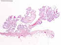
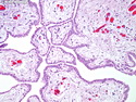
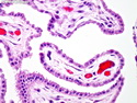
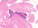
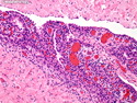
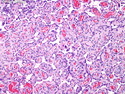
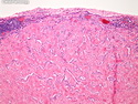
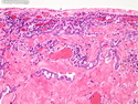
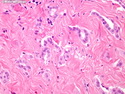
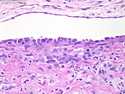
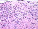
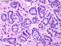
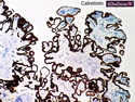
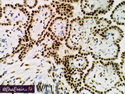
Mesothelial Hyperplasia
When taken out of context, mesothelial hyperplastic changes may be mistaken for mesothelioma or invasive carcinoma, especially when florid. Features favoring reactive hyperplasia include: absence of stromal invasion, uniform simple papillae lined by a single layer of mesothelial cells, inflammation, negativity for EMA, p53, GLUT-1, and IMP-3 markers, and positivity for desmin. Features favoring malignant mesothelioma over mesothelial hyperplasia include: grossly visible papillary lesions, stromal invasion with desmoplasia, dense cellularity, solid expansile nodules containing cells surrounded by stroma, complex papillae or tubules with cellular stratification, necrosis, positivity for EMA, p53, GLUT-1, and IMP-3 stains, and negativity for desmin.