
Testis : Anastomosing Hemangioma

slide 80 of 86


Comments:
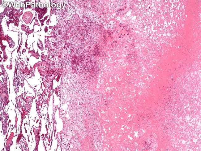
Anastomosing Hemangioma of Testis: Anastomosing sinusoidal vascular spaces can be seen in the left one-third of this image. The right two-thirds shows areas of thrombosis and necrosis. Some cases may also show foci of papillary endothelial hyperplasia and extramedullary hematopoiesis. The adjacent testicular parenchyma is usually atrophic without spermatogenesis.
slide 80 of 86