
Meningioma - Radiographic Features

slide 8 of 60


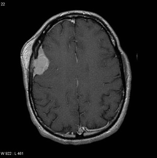
Comments:
MRI scan with contrast showing a meningioma extending through the skull vault to involve subcutaneous tissues. Despite the obvious atypical behavior, the tumor was well-differentiated and showed no atypia or necrosis. Case reproduced with permission, courtesy of Dr. Frank Gaillard; Radiopaedia; Complete case is here.
slide 8 of 60