
Type AB Thymoma : Microscopic

slide 8 of 20


Comments:
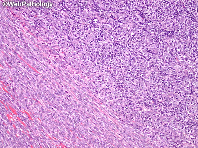
The lower left of this image of a type AB thymoma shows sheets of bland spindle epithelial cells (type A component). The upper right shows spindle cells admixed with numerous lymphocytes (type B component). The relative proportion of the two components is highly variable from case to case.
slide 8 of 20