
Hepatic Angiosarcoma : Clinical & Gross


Comments:
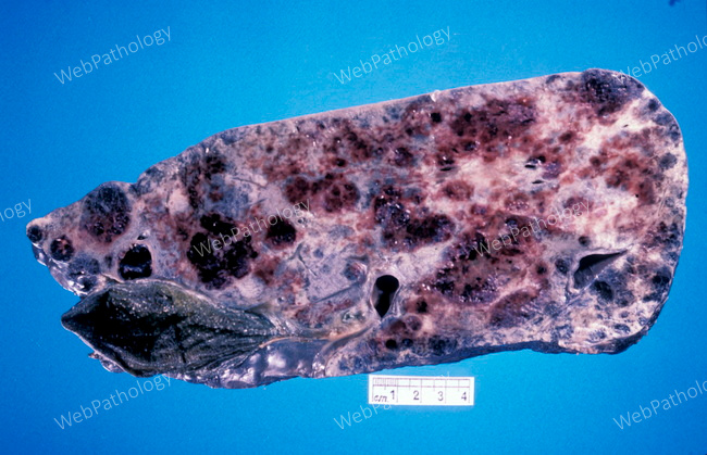
Clinical Features: Hepatic angiosarcoma usually occurs late in life, predominantly in the 6th and 7th decades. It is more common in men (M:F = 3:1). Patients present with vague symptoms and signs of liver disease and the diagnosis is often delayed. The clinical presentation consists of abdominal pain, fatigue, weight loss, anorexia, nausea, vomiting, abdominal mass, ascites, and jaundice. About 15%-20% of patients present to the ER with hemoperitoneum from tumor necrosis and rupture. This is considered to be a sign of malignancy because benign tumors rarely undergo spontaneous rupture. Rare cases present with acute liver failure. Many patients are asymptomatic and the tumor is discovered incidentally at autopsy. Patients may develop hematologic complications, including thrombocytopenia (secondary to platelet entrapment in the tumor), microangiopathic hemolytic anemia (due to RBC fragmentation in the tumor), and disseminated intravascular coagulation (due to local intravascular destruction of clotting factors). This specimen of hepatic angiosarcoma shows irregular, hemorrhagic and necrotic lesions scattered throughout the organ. Image courtesy of Dr. Ibrahim Zardawi; used with permission.