
Warthin's Tumor

slide 74 of 110


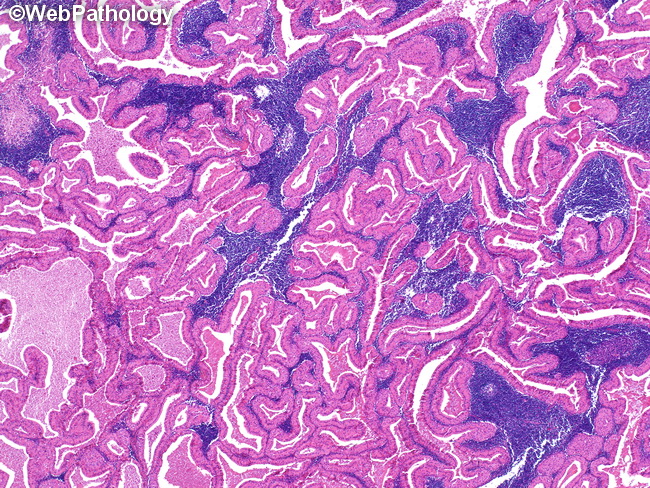
Comments:
Warthin�s tumor (WT) has a distinct morphologic appearance composed of papillary structures lined by bi-layered oncocytic epithelium forming cystic spaces and the stroma with lymphoid component (as seen in this low power micrograph). Other inflammatory cells such as plasma cells, mast cells, histiocytes and giant cells can be seen. Mucous cells and sebaceous glands can also be seen and their presence may raise the possibility of mucoepidermoid carcinoma.
slide 74 of 110