
Inflammatory Breast CA : Pathology


Comments:
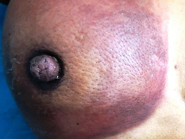
Inflammatory Breast Carcinoma (IBC) shows diffuse erythema and edema (peau d'orange) of the breast skin. By definition, there is involvement of at least a third or more of the skin overlying the breast (AJCC-TNM Staging System) and is classified as cT4d. Patients with characteristic changes but involvement of less than one-third of the skin are classified as cT4b or cT4c. IBC is primarily a clinical diagnosis and not a specific histologic subtype. The cutaneous changes of IBC are due to lymphedema caused by widespread carcinomatous emboli within dermal lymphatic channels. The tumor emboli are generally present throughout the breast but may or may not be seen in a small skin biopsy. Therefore, the presence of tumor emboli in dermal lymphatics is not required to make the diagnosis of IBC. Conversely, tumor emboli in dermal lymphatics in the absence of skin changes do not qualify as IBC. The diagnosis of IBC should not be applied to a patient with neglected locally advanced breast cancer that may be fungated or ulcerated. Pathologic Features: Grossly, the tumor is often indistinct in the mastectomy specimen. The entire breast is diffusely enlarged and indurated to due involvement by the tumor. The skin is visibly thickened. Microscopically, IBC is usually high-grade invasive ductal carcinoma of no specific type. IBC is characterized by negativity for ER and PR and overexpression of HER2. In addition, there is overexpression of p53, high E-cadherin expression, high MIB1 proliferation index, and cytoplasmic MUC-1 overexpression. There is prominent angiogenesis due to upregulation of angiogenesis-related genes. Bevacizumab - a recombinant monoclonal antibody to VEGF appears to have a role in the treatment of IBC. Most patients with IBC are treated by neoadjuvant chemotherapy followed by mastectomy and radiation. Case courtesy of: Dr. Sanjay D. Deshmukh (Professor of Pathology) and Dr. J. M. Gadekar (Chief of Surgery), Dr. Vithalrao Vikhe Patil Medical College & Hospitals, Ahmednagar, India