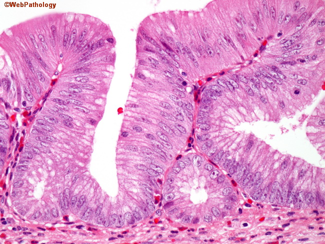
Mucinous Cystadenoma


Comments:
The image shows papillary structures lined by adenomatous epithelium in a mucinous cystadenoma of the appendix. The epithelium, while clearly atypical, still shows some order and low-grade cytology. Some mucinous tumors of the appendix are difficult to classify as clearly benign or clearly malignant. Tumors that are confined to the appendix may be histologically indistinguishable from those that have spread to the peritoneum causing pseudomyxoma peritonei. To address this conundrum, some authors subdivide mucinous tumors of the appendix into two groups: 1) Mucinous tumor of uncertain malignant potential OR Low-grade appendiceal mucinous neoplasm - this includes all lesions previously classified as mucinous cystadenomas as well as tumors that have spread to the peritoneum but without destructive invasion of the appendiceal wall; 2) Mucinous cystadenocarcinoma - which includes any appendiceal mucinous tumor with either high-grade cytology or destructive invasion of the appendix. Here is an excellent review of the subject: Appendiceal Mucinous Neoplasms: Controversial Issues. J Misdraji. Archives of Pathology & Laboratory Medicine: June 2010, Vol. 134, No.6, pp.864-870.


