
Toxoplasma Lymphadenitis
Home
 Hematopathology
Lymph Node (Non-Hematopoietic)
Protozoal Lymphadenitides
Toxoplasma Lymphadenitis
Hematopathology
Lymph Node (Non-Hematopoietic)
Protozoal Lymphadenitides
Toxoplasma Lymphadenitis

slide 6 of 7


Comments:
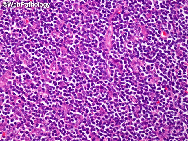
Toxoplasma Lymphadenitis: Monocytoid B-lymphocytes are one of the triad of histologic findings of toxoplasma lymphadenitis; the other two being hyperplastic follicles and epithelioid cell clusters. The lymphocytes are arranged in sheets within and around marginal and cortical sinuses and blood vessels. They are medium-to-large, uniform cells with pale or clear cytoplasm, and small hyperchromatic nuclei. Nucleoli are absent. Mitotic activity is not increased. They express B-cell markers and surface immunoglobulins. They are not specific for toxoplasmosis and are seen in other conditions such as HIV-associated lymphadenitis.
slide 6 of 7