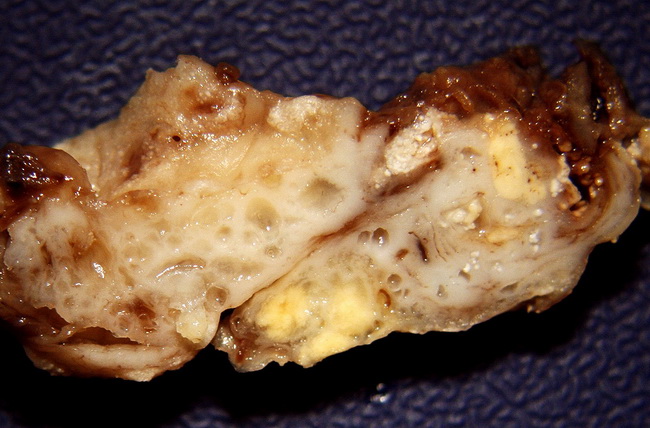
Adamantinomatous Craniopharyngioma


Comments:
Resected specimen of an adamantinomatous craniopharyngioma showing solid and cystic areas. The yellowish-white specks in the right half of the specimen represent the nodules of so called "wet keratin" composed of clusters of anucleated squames. Craniopharyngiomas show a bimodal distribution with the first peak around age 10-14 yrs. (mostly adamantinomatous type) and a second smaller peak around age 50-60 yrs. (mostly papillary type). Courtesy of: Dr. Luciano de Souza Queiroz, Dept. of Pathology, Faculty of Medical Sciences, State University of Campinas (UNICAMP), Campinas, Sao Paulo State, BRAZIL. Additional images are here.


