
Osteosarcoma : Radiographic Features


Comments:
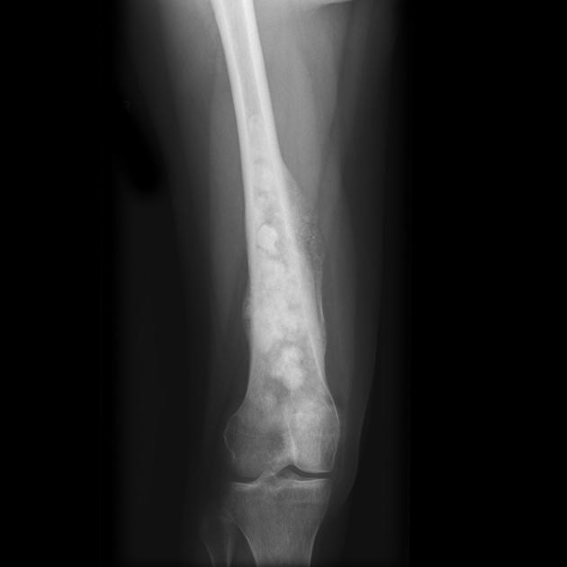
Osteosarcoma is a malignant tumor of bone where the tumor cells produce osteoid matrix. It is the 2nd most common malignant tumor of the bone (after multiple myeloma). The peak incidence is in 2nd decade. The sites most commonly involved are the metaphyseal region of long tubular bones, especially the region around knee joint. This plain radiograph (AP view) shows typical features of an osteosarcoma involving distal femur in a 17 y/o female. There is a destructive lesion with an admixture of lytic and sclerotic areas, elevation of periosteum by the proliferating tumor (Codman triangle), calcification of the tumor matrix, and a soft tissue mass. The osteoid matrix produced by the tumor cells often creates a "fluffy" or "cloud-like" appearance on plain films. Case produced with permission, courtesy of Dr. Frank Gaillard. Radiopaedia. Complete case is here.