
Fibrous Histiocytoma

slide 4 of 8


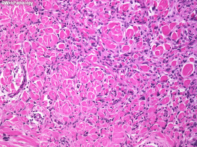
Comments:
Fibrous histiocytomas do not have sharply defined borders. They usually show entrapment of surrounding dermal collagen. Fibrous histiocytomas contain Factor XIII-a +ve cells and are negative for CD34. DFSP's lack significant number of Factor XIII-a +ve cells and express CD34 in the majority of tumor cells.
slide 4 of 8