Synovial Sarcoma : Break Apart FISH


Comments:
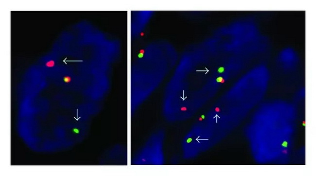
The balanced reciprocal translocation t(X;18)(p11.2;q11.2) which is diagnostic of synovial sarcomas can be detected with RT-PCR, FISH, or conventional cytogenetics. The currently preferred method is FISH analysis on formalin-fixed paraffin-embedded tissue using a dual-color break-apart SS18 probe. SS18 Dual Color Break Apart FISH Probe: The probe is available commercially and is designed to detect chromosomal translocations involving the 18q11.2 region that harbors the SS18 gene (aka SYT gene). The probe is a mixture of two direct labeled probes hybridizing to the 18q11.2 band. The green fluorochrome-labeled probe hybridizes proximal to the SS18 gene and orange fluorochrome-labeled probe targets the 18q11.2 region distal to the SS18 gene. Result Interpretation: Following hybridization, a normal interphase nucleus will show two green/orange fusion signals that represent two normal (non-rearranged) 18q11.2 loci. A cell containing 18q11.2 translocation will show one green/orange fusion signal (representing one non-rearranged 18q11.2 locus) and one separate orange signal and one separate green signal (indicating the translocation) as shown in this image. Reference (and image source): Yi-che Changchien et al. A Challenging Case of Metastatic Intra-Abdominal Synovial Sarcoma with Unusual Immunophenotype and Its Differential Diagnosis. Case Reports in Pathology, Volume 2012, Article ID 786083
[accessed 11 Dec, 2018] Used under license Creative Commons Attribution Unported (CC by 3.0)


