
Lipofibromatosis : Gross & Microscopic Features
Home
 Soft Tissue
Fibroblastic
Fibrous Tumors of Infancy
Lipofibromatosis : Gross & Microscopic Features
Soft Tissue
Fibroblastic
Fibrous Tumors of Infancy
Lipofibromatosis : Gross & Microscopic Features

slide 38 of 60


Comments:
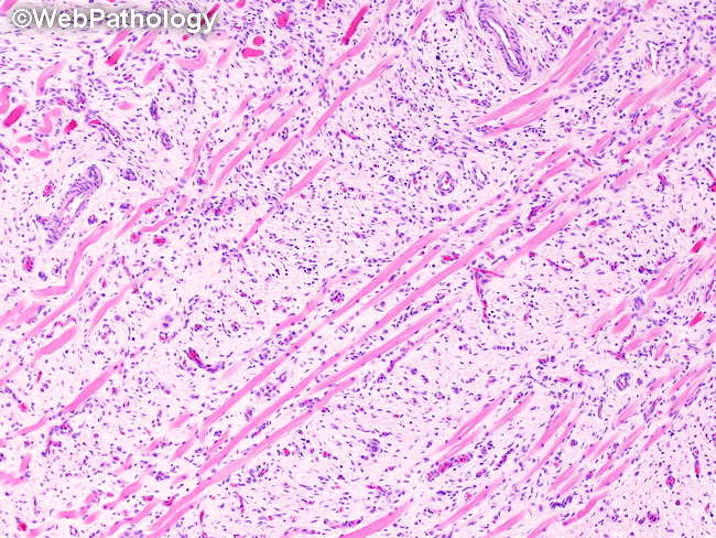
Gross Pathology of Lipofibromatosis: The resected specimen is a firm, gray to yellow-white, poorly-circumscribed mass ranging in size from 1 to 10 cm. Given its infiltrative nature, the wide local excision often includes surrounding skeletal muscles and subcutaneous fat. Microscopic Features: The early stages of lipofibromatosis (previously referred to as mesenchymal or diffuse pattern) consist of small round to oval primitive mesenchymal cells, haphazardly scattered in a myxoid matrix. The cells are intimately associated with adipose tissue (not seen in this image) and infiltrate between bundles of skeletal muscle fibers without destroying them and widely separating them as a result (as illustrated here).
slide 38 of 60