
Dedifferentiated Chordoma


Comments:
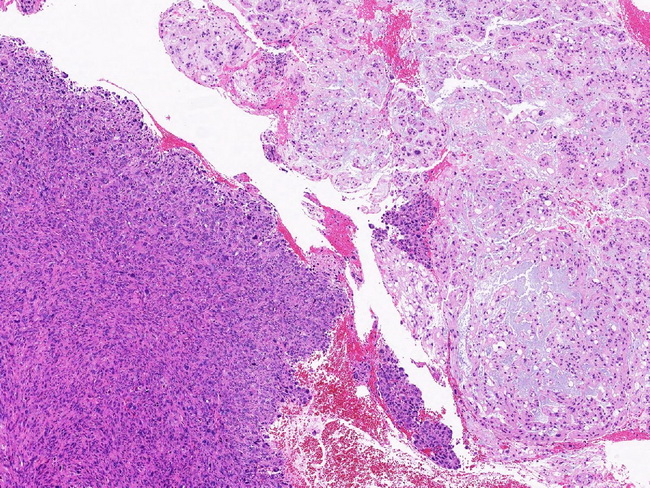
Dedifferentiated Chordoma: Dedifferentiated chordomas have a biphasic appearance consisting of high-grade sarcoma juxtaposed to conventional chordoma. They comprise <1% of all chordomas. They involve the usual sites along axial skeleton and can present de novo, post-radiation, or as recurrence/metastasis months to years after the initial presentation. Pain and neurological symptoms related to nerve compression are common. The prognosis is poor with a median survival of < 2 years. Grossly, dedifferentiated chordomas show gelatinous areas representing conventional chordoma adjacent to solid, fleshy, and pink-tan area representing the dedifferentiated component. Microscopically, the dedifferentiated component may be pleomorphic sarcoma, high-grade spindle cell sarcoma, osteosarcoma or rhabdomyosarcoma. By immunohistochemistry, the conventional chordoma is positive for brachyury and cytokeratin, whereas the dedifferentiated component lacks expression for both brachyury and cytokeratin and harbors TP53 mutations. Reference: Hung YP et al. Dedifferentiated Chordoma: Clinicopathologic and Molecular Characteristics With Integrative Analysis. Am J Surg Pathol. 2020 Sept; 44(9):1213-1223. Image courtesy of: John Gross, MD; used with permission.