
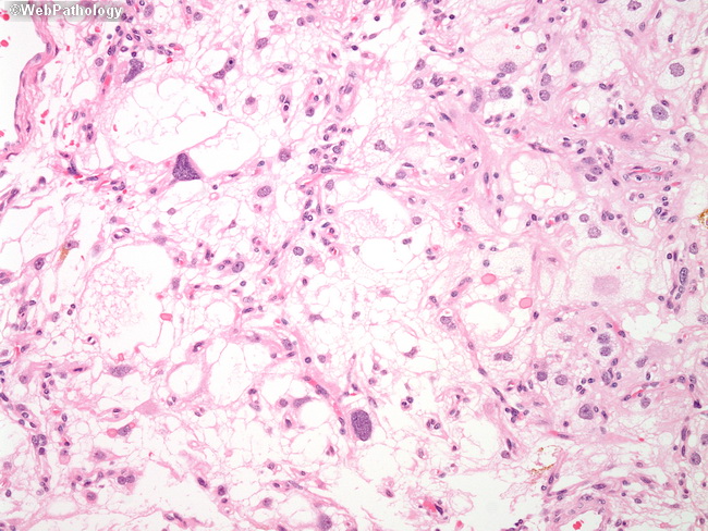
Microcystic Meningioma

slide 32 of 60


Comments:
High power view of the previous image. The majority of meningothelial cells are monotonous, displaying uniform nuclei with fine chromatin. At least two bizarre cells with prominent hyperchromatic nuclei are readily noted. It is a common finding in the microcystic variant.
slide 32 of 60