
Rhabdoid Tumor : Microscopic Features


Comments:
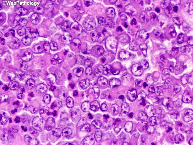
Microscopic Features: Rhabdoid tumor of kidney involves the medullary region and shows diffuse infiltration of the adjacent renal parenchyma with vascular invasion. The prototypic appearance consists of somewhat monomorphic population of high-grade malignant cells arranged in diffuse sheets. Other architectural patterns include solid, pseudoalveolar, trabecular, sclerosing, epithelioid, spindled, cystic, or myxoid patterns. The tumor cells have indistinct cytoplasmic borders and a large vesicular nucleus with 1-2 prominent nucleoli. One of the characteristic features is the presence of large, intracytoplasmic eosinophilic hyaline globule composed of intermediate filaments which causes lateral displacement of the nucleus and imparts a plasmacytoid or rhabdomyosarcomatous appearance.