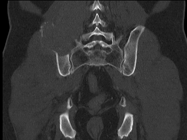
Solitary Plasmacytoma : Bone


Comments:
Solitary plasmacytomas make up 3% to 5% of plasma cell neoplasms. The median age at presentation is about 55 years which is 10 years younger than that for multiple myeloma. There is a strong male predominance. Most patients present with a solitary painful bony lesion or with a pathologic fracture. Some are asymptomatic and discovered incidentally. The radiologic features of lytic bone lesion are similar to those of multiple myeloma. Given the most commonly involved location (thoracic spine), some patients present with features of spinal cord or nerve root compression. This CT scan of pelvis (coronal bone window) is from a 65 y/o male with long-standing history of low back pain. There was no recent trauma. The CT scan shows a lytic soft tissue expansile lesion of the superior iliac wing, adjacent to the sacro-iliac joint. It contains foci of calcification. It partly destroys the right L3-L5 transverse processes. The diagnosis of plasmacytoma was confirmed on a CT-guided biopsy. Case courtesy of Dr Henry Knipe, Radiopaedia.org. From the case rID: 42879


