
Chondroid Chordoma


Comments:
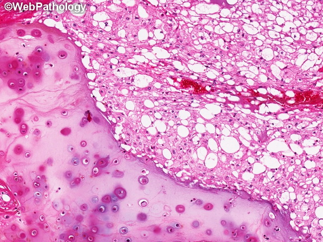
Chondroid chordoma is a variant that almost always arises at the base of the skull and has features of both conventional chordoma (right) as well as chondrosarcoma (left). The transition between conventional and chondroid areas may be abrupt (shown here) or gradual. The chondroid areas show neoplastic cells in lacunar-like spaces embedded within basophilic matrix resembling neoplastic hyaline cartilage. When chondroid component predominates, such cases may be difficult to distinguish from chondrosarcoma on morphology alone. An immunohistochemical panel of brachyury, cytokeratin, EMA, and S100 is sufficient in resolving most cases. Chordomas are positive for all four markers, where as conventional chondrosarcomas are brachyury-, cytokerain-, EMA- and S100+. Extraskeletal myxoid chondrosarcomas are brachyury-, cytokeratin-, EMA+ and S100-. In addition, they show NR4A3 rearrangement. The prognosis of chondroid chordomas is similar to that of conventional chordomas.