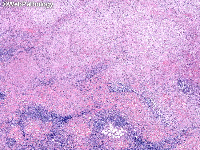
Extranodal Rosai-Dorfman Disease : Skin


Comments:
The skin lesions of cutaneous Rosai-Dorfman disease show a dense cellular infiltrate of histiocytes and lymphocytes in the dermis and subcutis. In addition, there may be plasma cells, neutrophils, and eosinophils. The infiltrate may have a nodular pattern (as seen here). Some cases show stromal fibrosis with a storiform pattern. Emperipolesis is not conspicuous. Ulceration, necrosis, cytologic atypia, increased mitotic activity, and granulomas are not present. Rare cases may show reactive lymphoid follicles.


