
Neurofibroma of Bladder

slide 22 of 28


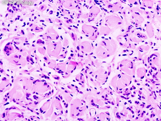
Comments:
Higher magnification of the previous image showing Wagner-Meissner corpuscles. They are separated by spindle cells with oval or elongated nuclei. The tumor may show diffuse and/or plexiform growth patterns.
slide 22 of 28