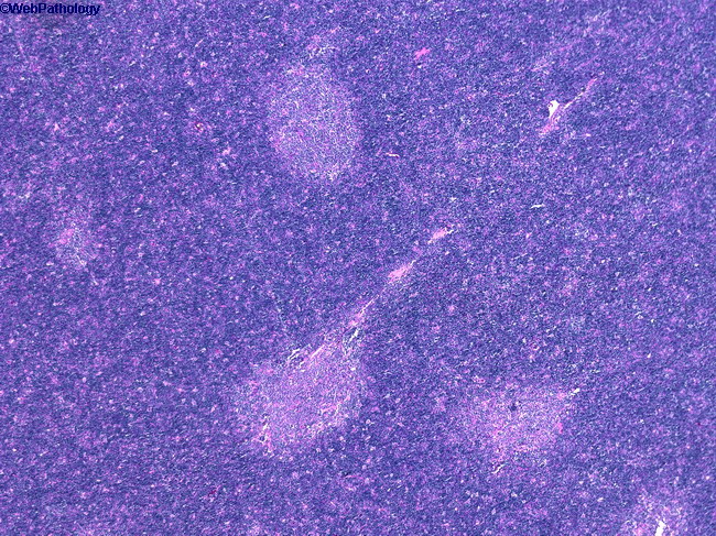
Type B1 Thymoma (Lymphocyte-rich) : Presentation


Comments:
Type B1 thymoma accounts for about 18% of all thymomas. It is seen most commonly in the 5th and 6th decades of life with a slight female predominance. Two-thirds of patients are symptomatic and present with symptoms related to compression of anterior mediastinal structures (cough, chest pain, dyspnea) or autoimmune diseases (myasthenia gravis, pure red cell aplasia, hypogammaglobulinemia). Between 20% to 50% have myasthenia gravis. Almost 90% of cases are either completely encapsulated grossly and microscopically (Stage I) or show only microscopic infiltration of the capsule (Stage II). Extension to adjacent mediastinal structures is rare. The is a low power view of Type B1 thymoma showing a predominance of dark blue cortical areas and a few pale islands of medullary differentiation. The cortical areas are composed of scattered neoplastic epithelial cells in a background of a dense infiltrate of immature T-cells. The paler medullary areas are less cellular and contain few or no immature T-cells but have increased numbers of B cells and mature T-cells, and may have myoid cells and Hassall corpuscles.


