
Kikuchi Lymphadenitis : Microscopic
Home
 Hematopathology
Lymph Node (Non-Hematopoietic)
Lymphadenopathies - I
Kikuchi Lymphadenitis : Microscopic
Hematopathology
Lymph Node (Non-Hematopoietic)
Lymphadenopathies - I
Kikuchi Lymphadenitis : Microscopic

slide 17 of 69


Comments:
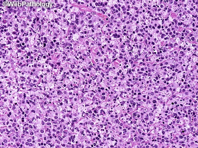
The predominant lesional cells in Kikuchi lymphadenitis are histiocytes, plasmacytoid dendritic cells, and activated T-cells. Histiocytes often show crescentic nuclei and contain phagocytized debris (as seen here). They are CD68+, CD163+, and contain cytoplasmic myeloperoxidase. The plasmacytoid dendritic cells are CD123+ and TCL1+ and cluster at the periphery of necrotic lesions.
slide 17 of 69