
Cardiac Amyloidosis : Thioflavin T

slide 16 of 22


Comments:
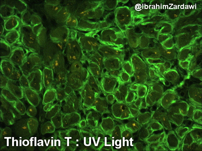
Cardiac Amyloidosis: This image shows amyloid deposits in perimyocytic distribution stained with Thioflavin T and visualized under ultraviolet light. Image courtesy of: Dr. Ibrahim Zardawi; used with permission.
slide 16 of 22