
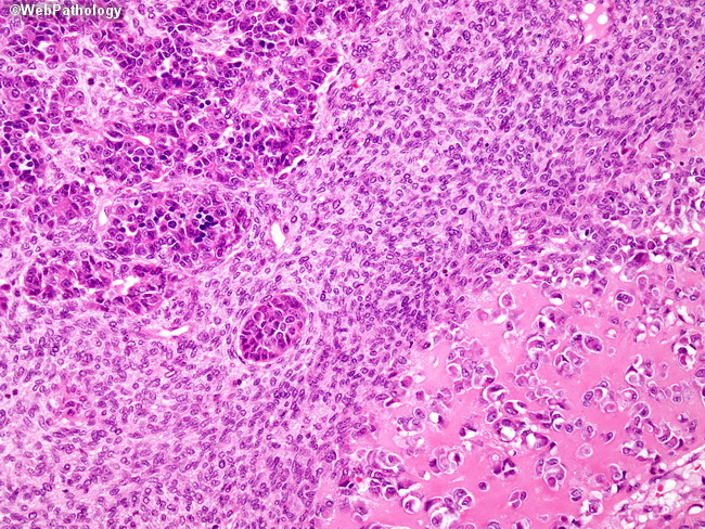
Hepatoblastoma - Mixed Pattern

slide 16 of 65


Comments:
As in the previous image, this mixed epithelial-mesenchymal hepatoblastoma contains both embryonal epithelial-type differentiation (top left) and osteoid matrix (bottom right), which are separated by bands of primitive mesenchyme (numerous plump spindle cells). Foci of hematopoietic precursors, representing extramedullary hematopoeisis, may be seen in the epithelial areas.
slide 16 of 65