
Hepatoblastoma - Mixed Pattern

slide 14 of 65


Comments:
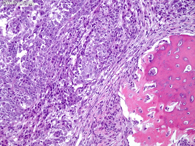
Higher magnification of this mixed epithelial-mesenchymal hepatoblastoma shows both embryonal epithelial-type morphology (left) and osteoid matrix (right). Note the high nuclear:cytoplasmic ratios and marked cellular atypia in the embryonal area. The epithelial and mesenchymal elements are separated by primitive mesenchyme, which comprises undifferentiated spindle cells. Following chemotherapy, the epithelial areas are typically necrotic and only mesenchymal components may be identified.
slide 14 of 65