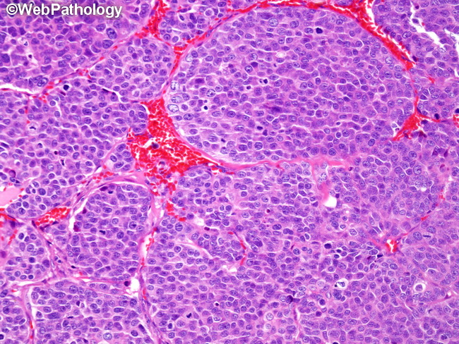
Dedifferentiated Adenoid Cystic CA


Comments:
The image shows a focus of de-differentiated adenoid cystic carcinoma. The patient reported sudden increase in the size of a long-standing tumor in the submandibular region. The tumor was composed of solid sheets of pleomorphic cells with central necrosis. Mitotic activity was brisk (see next two images). Areas of conventional adenoid cystic carcinoma with tubular architecture were present in the periphery of the tumor.


