
Cardiac Myxoma : Microscopic Features

slide 13 of 102


Comments:
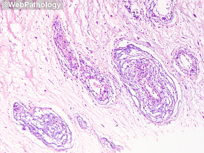
Microscopic Features: Histologically, cardiac myxomas are composed of nests and cords of myxoma cells (aka lepidic cells) in a myxoid background. The cells are round, oval, polygonal, fusiform or stellate shaped with bland nuclei, punctate nucleoli and light pink cytoplasm. Multinucleated tumor cells may be seen.The cells may be found singly as well as in small nests, syncytial cords, or tubular structures. They are closely associated with and cluster around vascular channels, forming one or more concentric layers or rings around them.
slide 13 of 102