
Fibrous Hamartoma of Infancy : Differential


Comments:
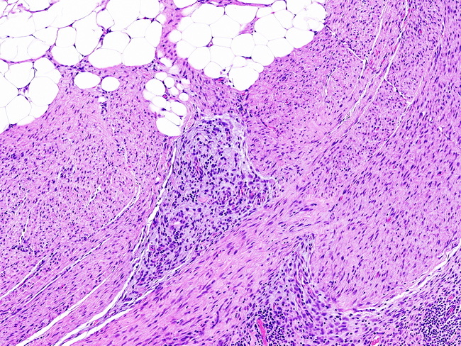
Differential Diagnosis of Fibrous Hamartoma of Infancy (FHI) (continued from the previous image): For FHI with a dominant fibroblastic component, the differential diagnosis includes: Desmoid-type Fibromatosis: Rare in young children; long, sweeping fascicles of band fibroblastic cells; nuclear staining with β-catenin; mutations of CTNNB1 gene. Diffuse Myofibromatosis: Consists of hemangiopericytomatous vascular spaces separating lightly-staining nodules. Calcifying Aponeurotic Fibroma: Located in distal portion of the extremities in older children; calcified and chondroid-appearing nodules surrounded by epithelioid cells; presence of FN1-EGF fusion. The image shows classic triphasic appearance of FHI. The differential diagnosis continues in the next image.