
Radiation Necrosis (Osteoradionecrosis) : Introduction


Comments:
Necrosis of bone in the radiation field is a known late complication of radiation therapy. It can involve any bone, but the most commonly affected sites are jaw bones, ribs, pelvis, spine, and humerus. Necrosis begins usually within 3 years of radiation therapy but may be delayed by decades. Patients are usually older than 55 years of age.
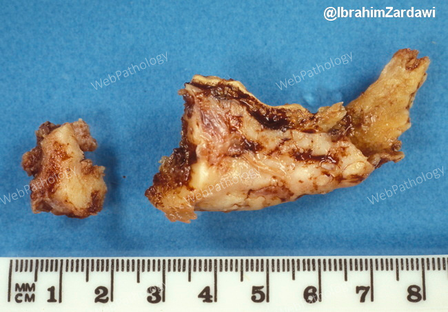
There is approximately 9% incidence of developing mandibular osteoradionecrosis in patients receiving >7000 centrigrays of radiation for head and neck cancers. The risk of this complication has decreased with newer modalities such as intensity-modulated radiation therapy. This partial mandibulectomy specimen shows pathologic fracture secondary to radiation necrosis. Image courtesy of: Dr. Ibrahim Zardawi; used with permission.